Concurrent MRS during tACS or other neuromodulation: meaningful online effects, and how severe are artifacts?

Hi everyone,
I’m planning an MRS study combined with noninvasive neuromodulation (e.g., tACS), and I’d really appreciate advice from anyone with experience or relevant references.

I have two main questions:

1. Is it meaningful to acquire MRS during stimulation (online MRS)?
For neurotransmitters/metabolites (e.g., GABA, Glu/Glx), do changes typically appear more reliably as offline/aftereffects rather than during the stimulation period? In other words, is “mid-stimulation MRS” expected to capture physiologically interpretable changes, or is it often dominated by non-specific factors (state changes, attention/arousal, etc.) and low sensitivity?
And although I’ve read some articles found the online effect by MRS(mostly tDCS), I still wonder whether neuromodulation can have effect on neurotransmitters in that short time and while MRS can capture that change.
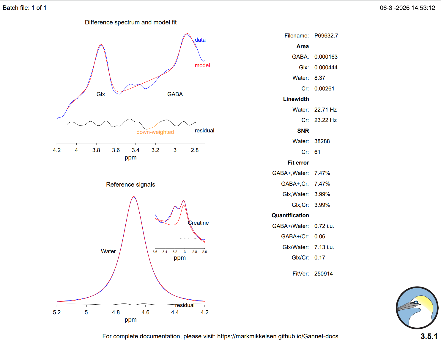
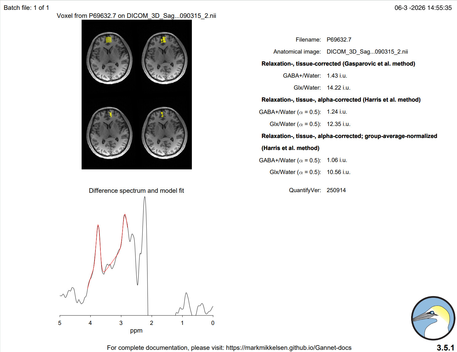
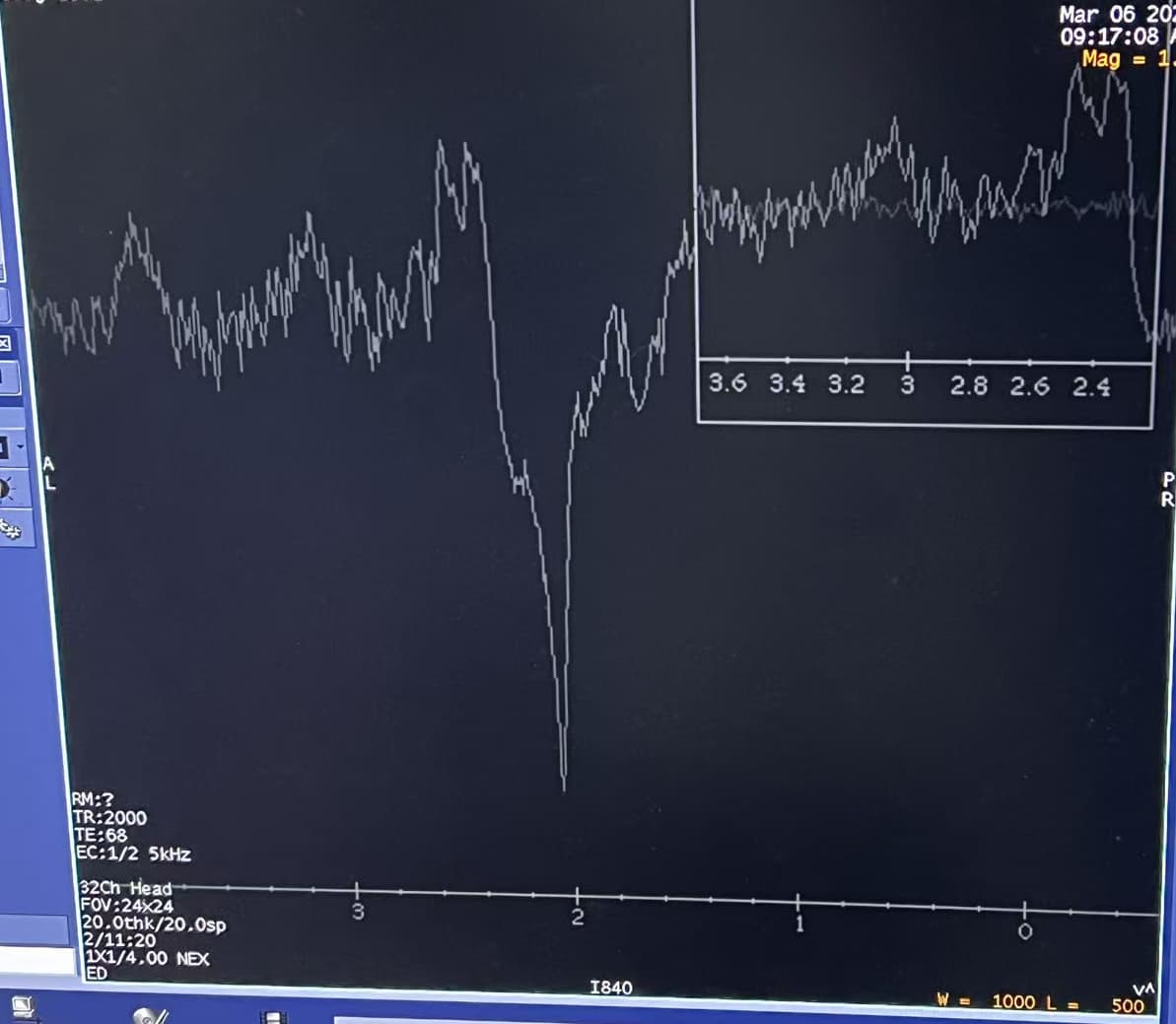
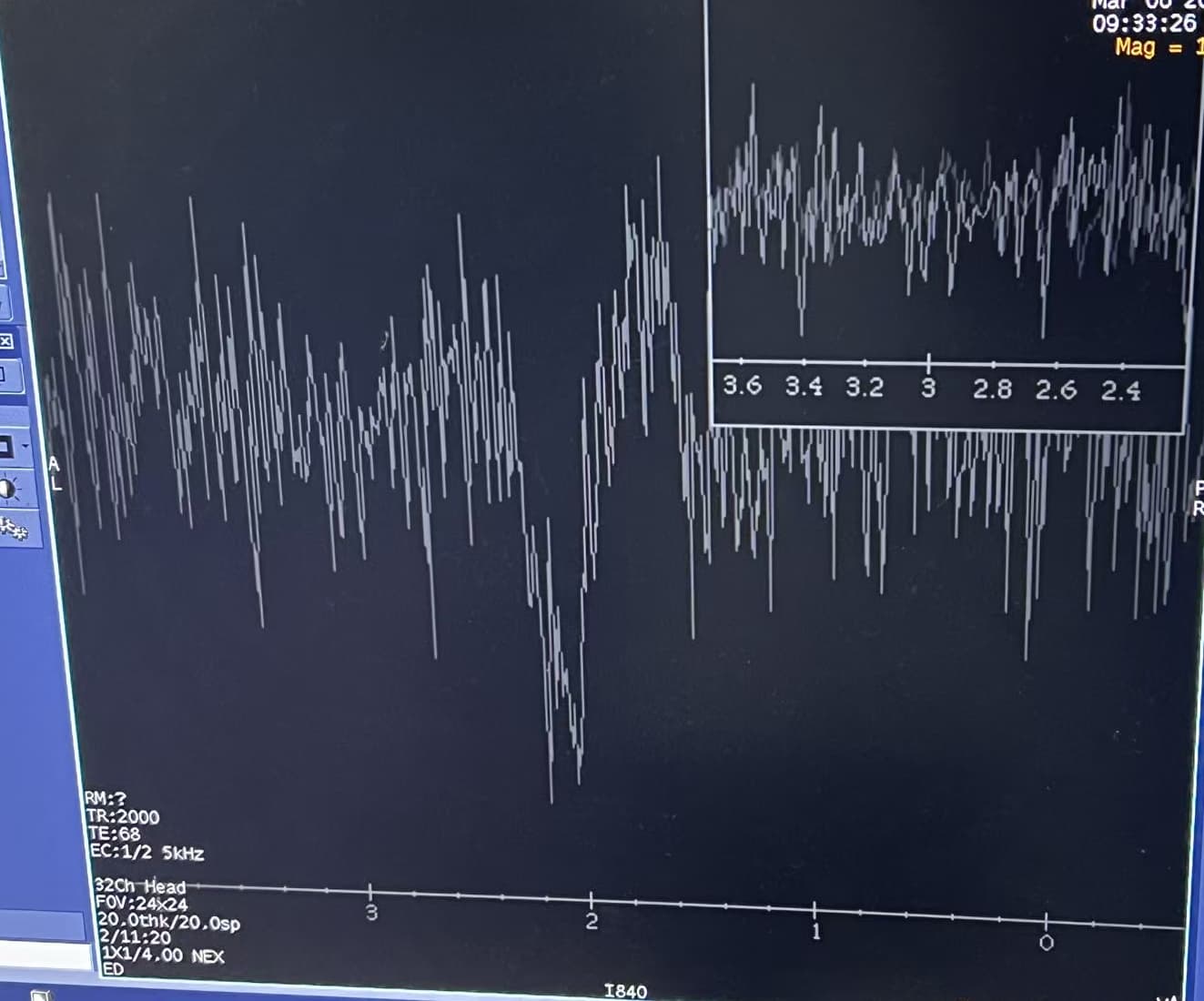
2. How much does stimulation hardware/current affect MRS data quality (artifacts)?
For example, with high-frequency currents or typical tACS setups, do you see substantial MRS artifacts (RF interference, line noise, spurious peaks, frequency/phase instability, B0 drift, degraded linewidth/SNR, etc.)?
I’m aware that concurrent stimulation can strongly affect BOLD/fMRI signals—does something similarly problematic happen for MRS, or is it generally manageable with proper setup (cable routing, filtering, synchronization, pausing stimulation during readout, etc.)?

If you have practical recommendations (sequence choices, QC metrics to watch, whether to interleave ON/OFF blocks, filtering/shielding strategies, vendors/devices that work better, or “don’t do it” warnings), I’d be very grateful.

Thanks in advance!

Hi @jmy_psy,

I asked these questions to the PiNG group at Oxford. Poly kindly gave these answers.

Regarding question 1:

The time-course of NIBS effects depends on a lot of factors, included, but not limited to, the exact stimulation protocol (intensity, duration, waveform, e.g. here) and brain state at the time of stimulation.

Previous work using tDCs has shown changes occurring during stimulation but changes (esp. in GABA) are often more pronounced following stimulation (e.g. Modulation of GABA and resting state functional connectivity by transcranial direct current stimulation | eLife). It is worth considering that, due to homeostatic plasticity, cortical excitability does eventually return to baseline, sometimes overshooting its previous state (i.e. if using an inhibitory protocol, after some time the inhibited tissue may become shortly excited), thus if looking at offline effects only, the timing of the measurement matters. Additionally, the majority of the literature investigate the mechanistic effects of tDCs is in the motor and visual cortex, which doesn’t necessarily mean it will translate to any cortical region in the same way.

tACS has not been studied as extensively in combination with MRS, thus the expected time-course is less clear.

Regarding question 2:

We have not observed any artefacts across scanner sites and protocols, with tDCS or tACS. However, we’ve always done temperature testing and pilots to ensure this isn’t the case before every new study using the equipment and piloting the exact stimulation protocol.

Hope this helps.

Will

Hi Will,

Thank you very much for taking the time to ask the PiNG group about my questions. I really appreciate your help with this. Please also extend my thanks to Dr.Poly and the PiNG team for their detailed and thoughtful responses.

Their explanations regarding the time-course of NIBS effects and the potential role of homeostatic plasticity are very helpful for us to reconsider our design.

Many thanks again
Best regards,
Jmy

Hi @jmy_psy, @wclarke ,

I can basically second this; in a small fraction of cases, we were able to observe very subtle RF interference during ramp phases of tDCS, but this was outside the typical spectral range and barely above background noise level. Definitely worth piloting each study to be sure though.

Hello everyone,

I recently conducted a pilot experiment using MEGA-PRESS MRS on a GE Discovery MR750 3T scanner. The experiment includes three stages: pre-stimulation, during stimulation, and post-stimulation. After processing the data with Gannet, I noticed several issues related to the reference spectra, linewidth, fitting errors, and noise, and I would like to ask for advice on what might be causing these problems and how they might be improved.

1. Abnormal reference signals (Water and Creatine)

In all three stages, the reference signals (Water and Creatine) show some abnormalities. As shown in the figure, the left panel shows the data from the pilot experiment.


The Water and Creatine peaks in our data partially overlap, whereas they are clearly separated in the normal data.

This makes me wonder whether the issue could be related to:

  • poor B0 shimming
  • scanner settings
  • signal-to-noise ratio
  • or possible interference from experimental equipment (e.g., electrode cables)

2.Question about the exponential line-broadening parameter (LB) in Gannet
In the Gannet processing pipeline I also modified the following parameter:
MRS_struct.p.LB = 3; % Exponential line-broadening (in Hz); default = 3 Hz
During the scan the operator mentioned that for GE systems this parameter might need to be adjusted depending on the actual linewidth observed in the data, and our measured linewidth values are already around 12–13 Hz, I wonder if this mean that we have low SNR here.

3. High-frequency noise during the stimulation stage
Another issue appears during the stimulation stage. Compared with the pre- and post-stimulation spectra, the spectrum acquired during stimulation shows clear high-frequency fluctuations across the spectrum. We can see it from the second picture comparing it to the first one.


When processed using Gannet, this stage also produces much larger fit errors compared to the other two stages.

I would like to ask whether this type of high-frequency noise during stimulation experiments is commonly observed in MRS studies and what strategies are typically used to reduce it.

Any suggestions regarding possible causes, acquisition improvements, or processing strategies would be greatly appreciated.Also, if anyone notices other potential issues in the data, I would be very grateful for any feedback. I still feel there may be other problems that I am not fully aware of, such as whether the ROI size or placement is appropriate, or other factors affecting the data quality.

Thank you very much for your help.

Hi @jmy_psy,

I can’t speak to the stimulation-related artifacts you may be observing, as I don’t have experience with concurrent stimulation-MRS experiments, but I can say that the shimming is quite poor. This is evident from your spectra and the water/Cr linewidths (>20 Hz). Ideally, you want them to be <~12 Hz.

Regarding Gannet’s default exponential line-broadening: yes, with poorly shimmed data, 3 Hz will exacerbate things. In my opinion, there is no recommended value for line-broadening, so I would try reducing the set value to 1.5 or 2 Hz. That said, it won’t address the shimming problem. Prefrontal voxels are very difficult to shim well. One possible recourse would be dielectric padding, but I’ve never attempted this, and you would, of course, need to have the equipment available.

Could you also post the GannetLoad output? My other concern is how well the frequency alignment is working, given the large linewidth of the data.

Mark

P.S. I don’t know if Glx is another target of interest, but I would not trust the Glx quantification value(s), because the model is fitting the left-hand shoulder of the Glx peak (as it expects a double-Lorentzian peak).

Hi @mmikkel ,

Thanks so much for your kindly answers. The following three pictures show the gannetload results for pre-modulation, during-modulation, and post-modulation, respectively.


I will try to redo this experiment using a shielding board or cancel the data collection during the stimulation phase, because the electrodes and EEG cap have indeed had a significant impact on the uniform field. Moreover, MRS seems to be more sensitive to the shimming than BOLD.

Ah, I see that you’ve set the line-broadening to 12 Hz. I misunderstood. This is uncharacteristically high and will lead to even large linewidths. What does the data look like if you leave the setting at 3 Hz?

It seems that the fit is worse with line-broadening set to 3 Hz, I was told to change the line-broadening to the value that GE has before every scanning begins, but I’m not entirely clear on the principles behind this adjustment. However, the quality of this data may indeed be problematic. I’m uncertain whether the equipment (such as the EEG cap and electrode wires) has affected the uniformity of the magnetic field, especially since there are issues with data quality even when no stimulation is applied.

Pre-modulation:


During-modulation:


Post-modulation:


Actually, the data look much better with the 3-Hz default, except for the during-modulation spectrum and the fit of the post-modulation spectrum. But that is less about shimming and more about noise/possible RF interference. I will leave it to the EEG-MRS users to comment on that.