I am wondering if anyone knows the physics behind why some of the lipid/MM peaks in MRS are inverted?
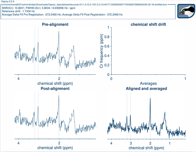
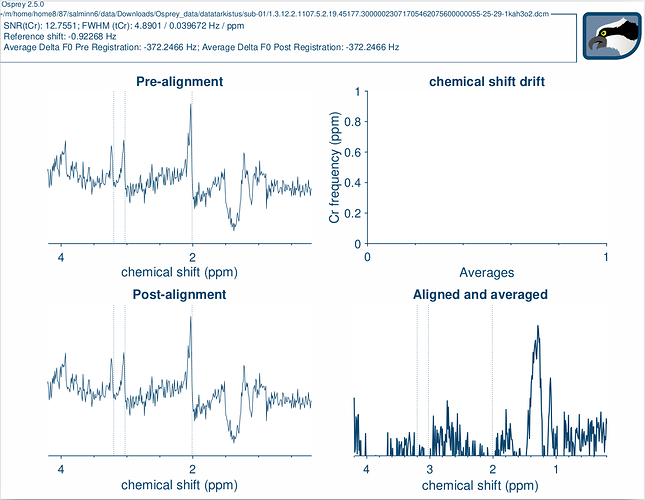
Here is an example: both of these spectra were taken from the same person in the same PRESS sequence. (concentrate on the post-alignment images)
I know that lactate can show as an inverted couplet at a specific TE (~144ms) is this the same for lipids/MMs?
I used a TE of 40ms on a Siemens Skyra 3T magnet.
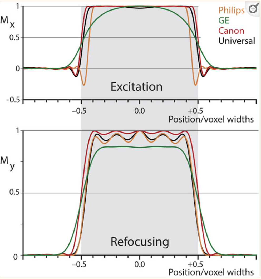
I have seen this happen with Philips data, and I’ve convinced myself that it can be caused by small side lobes of the excitation pulse profile. At least on Philips, we know that the phase of these side lobes is negative. If the slice-selective gradient directions and voxel placement just happen to be right (or well, wrong), the lipids are hit smack by the anti-phased side lobe.
I don’t recall the Siemens profile (and we weren’t allowed to show it in that paper), but I wouldn’t be surprised if there is a similar negative dip. I don’t think this could be a J-modulation thing at 40 ms.
Hi,
where are both voxels located and did you use outer volume suppression? I can also imagine that it’s a combination from outer voxel contamination and B0 inhomogeneities, so that the lipid signal isn’t refocused from the 180° pulses and has the right phase from the inhomogeneities.
Best,
Heiner
And now that you mentioned outer volume suppression. That could be the problem. I hadn’t researched that before and I believe that the scanner didn’t do that on default.
If you’re using the stock Siemens sequence, there is no outer volume suppression (like in the Minnesota sequence), although you can add a couple of saturation bands around the voxel.
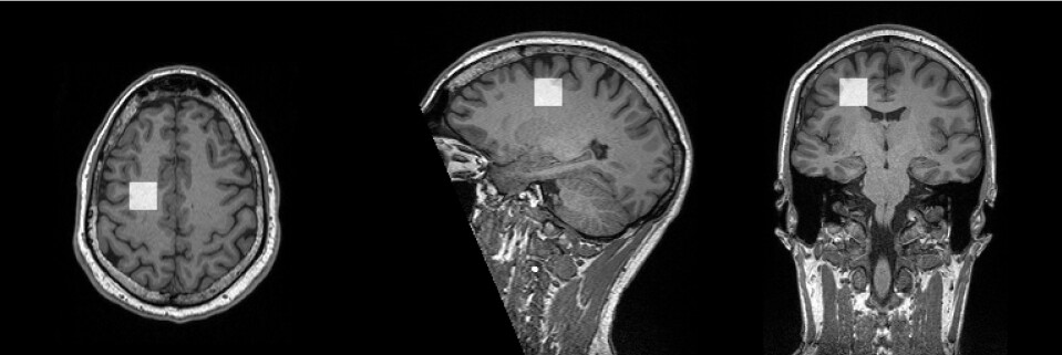
This one seems a little too far away from the edge of the brain to be contaminated by lipids in the first place. Do these participants tend to move more than others?
Finally, having seen a couple of your spectra now, they seem very low-SNR. What are you trying to measure/accomplish?
I am using a stock Siemens sequence, but it is for MRS. Also I just learnt about the saturation bands, so I will try to use those in the future.
My participants are pretty good at staying still, since they are my colleagues and they know how the magnet works.
I am trying to see if it is possible to conduct a reliable fMRS study with our 3T scanner. So in this case we had the participant close their eyes and open them and see if there is a change in Glu, Gln or Glx.
In this particular case I also wanted to see what the spectra looked separately, but usually when I have multiple spectra and I average them I get SNR of 35-50 which is still quite low. I have decided to make the voxel a little bigger for my next scan, but do you have any other recomendations for improving the SNR?
Hi Nella,
have I understood it right that the two spectra are from one measurement? When you can exclude movement, you have to take the phase cycle into account. In a phase cycle, the lipid signals from outside the voxel can also alternate (and in one phase cycle the lipid signal will be cancelled out).
Maybe this sequence can also be interesting for you:
Not a lot, other than making sure that the shim is as good as it gets. Not to discourage you, but fMRS is hard. Have you done ‘conventional’ MRS before?
Thank you for all of your help.
I have been working on a little bit of everything. So I mixed up here. That particular voxel placement was just to see how the different voxel locations change the SNR and how close to the scalp do we have to be. The eyes-open eyes-closed was done at the visual cortex.
The experimantal design for the visual cortex experiment was very simple. I had my participant in the scanner and first took a baseline measurement (50 averages) and water reference data, then I asked the participant to consentrate on a cross with eyes open and again took the 50averages, then I asked them to close their eyes (50 ave), concentrate on the cross again (50) and lastly again close (50). At that point I was and am still very new to fMRS, so I didn’t know how many averages you needed for a good spectra and I have now changed my design a lot. Now I usually take 4-8 timepoints from each scenario and each timepoint has 16 spectra to average.
I hope you understood from my explanation, English is not my first language.