Our group proposes a 31-P MRSI sequence utilizing Rosette-Petal shaped 3D k-space acquisition to achieve 1 mL isotropic resolution (10x10x10mm^3) over a FOV of 480x480x480mm^3 with a total scanning duration of 6.7 minutes using a Siemens Prisma 3T/7T scanner.
Method
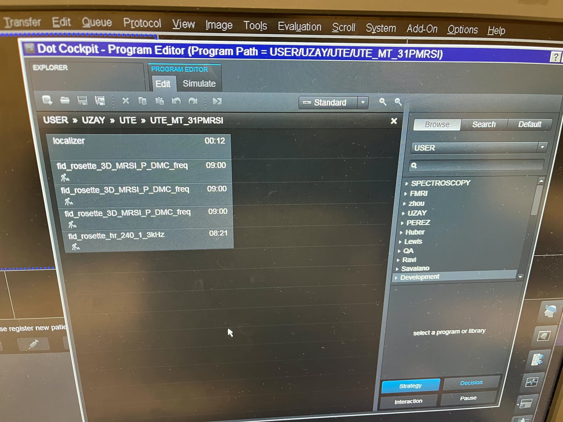
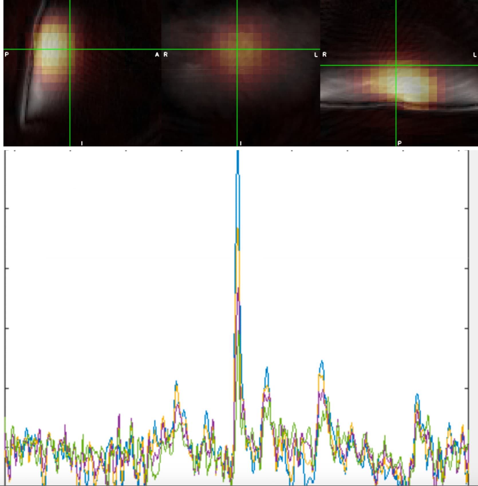
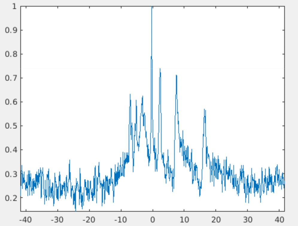
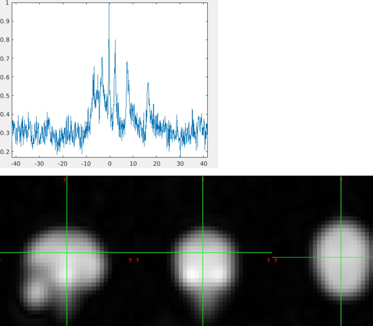
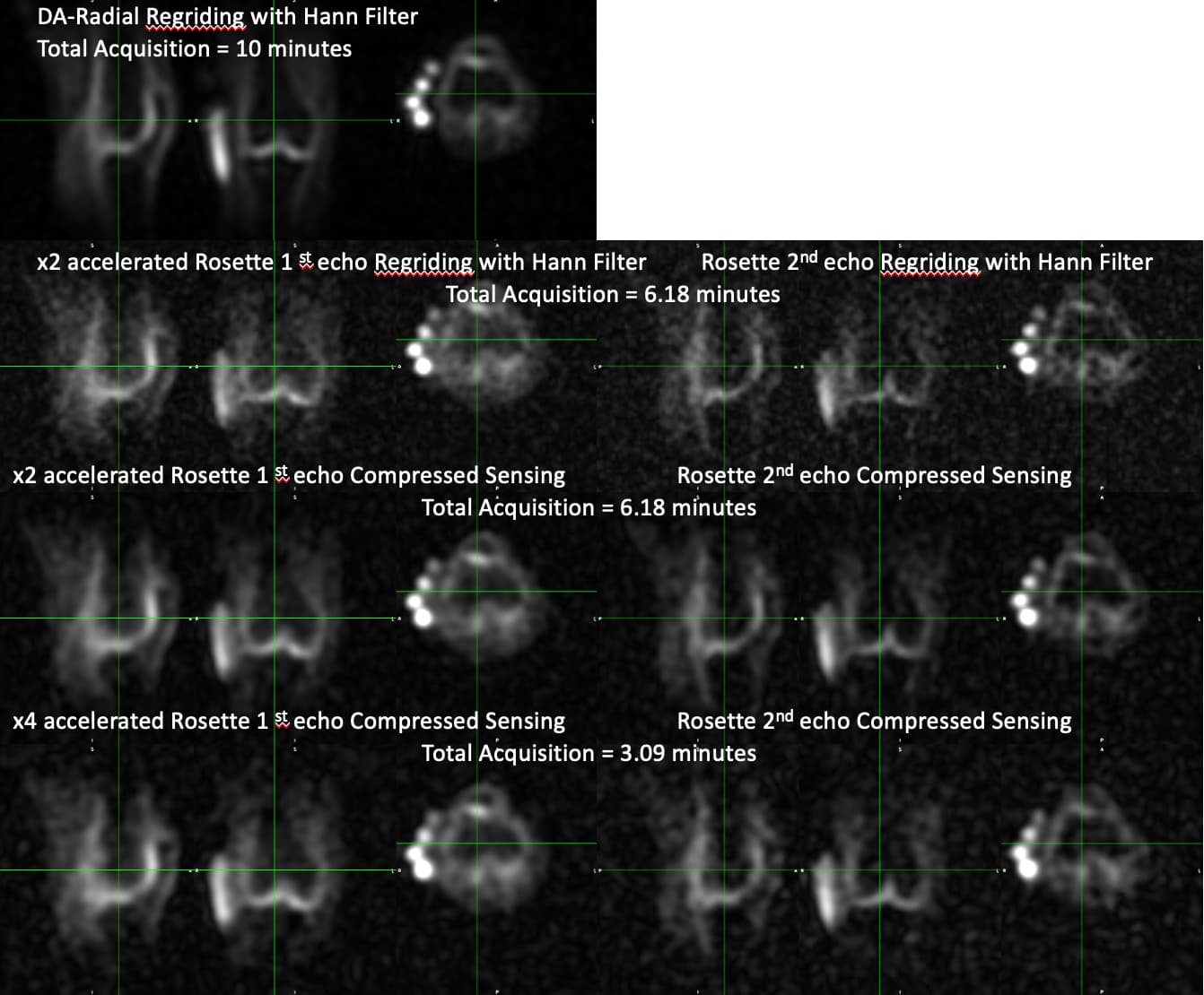
A custom rosette trajectory (Shen et al. 2022) was used with an isotropic FOV = (480mm)^3 and an imaging matrix grid of 48x48x48 voxels. The spectral bandwidth was 2083 Hz, a TE = 50 us, a TR = 350 ms, and a total of 1444 Petals. Total scan duration = 8.4 minutes. Anatomical data was acquired through an Ultrashort Echo Time Magnetization Transfer (UTE-MT) sequence which uses a pair of adiabatic hyperbolic secant (sech) 180-degree pulses with a pulse duration of 24ms, a pulse bandwidth of 1kHz, an offset frequency of ~1300 Hz from water. Image reconstruction and post-processing steps were performed in MATLAB (MathWorks, USA). Nonuniform fast Fourier transform (NUFFT) was used to calculate the forward encoding transform of the acquired k-space data. A compressed sensing approach was used for image reconstruction, using total generalized variation (TGV) as the sparsifying penalty. The complex-valued coil sensitivity maps were first extracted from the center of k-space using ESPIRIT for coil combination. We used LCModel for the quantification of in vivo 31P NMR Brain Spectra.
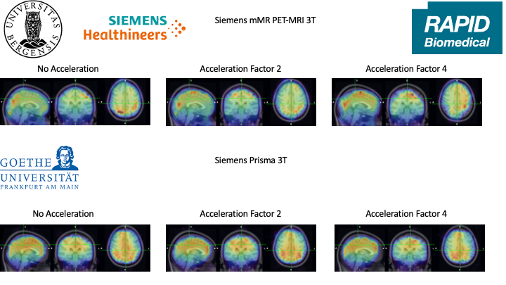
In order to achieve acceptable acquisition timing in clinical settings (reconstructed images with acceleration factors = 2 and 4 were tested for further acceleration purposes.
Shen, Xin, et al. “Ultra-short T2 components imaging of the whole brain using 3D dual-echo UTE MRI with rosette k-space pattern.” Magnetic Resonance in Medicine (2022).
Here is the proposed acquisition protocol… The Total acquisition duration will be 36 minutes. We will test the acquisition with an acceleration factor of up to 6 corresponding to a 31P MRSI acquisition of 4.5 minutes.
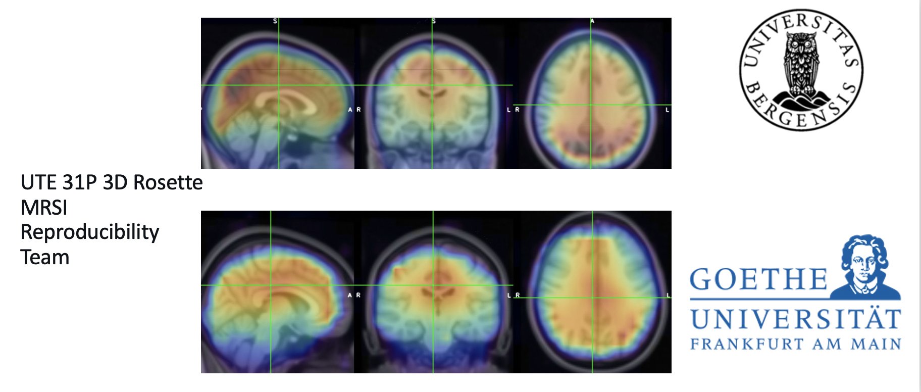
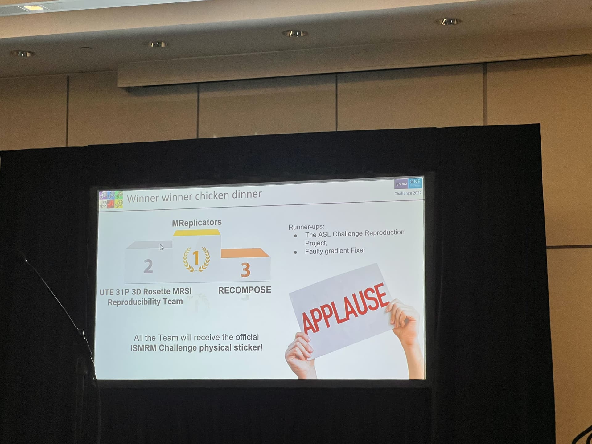
UTE 31P 3D Rosette MRSI Reproducibility Team is on board.
uzay emir Purdue University
Ulrich Pilatus Goethe University Frankfurt
Seyma Alcicek Goethe University Frankfurt
Alexander Richard Craig-Craven UNIVERSITY OF BERGEN
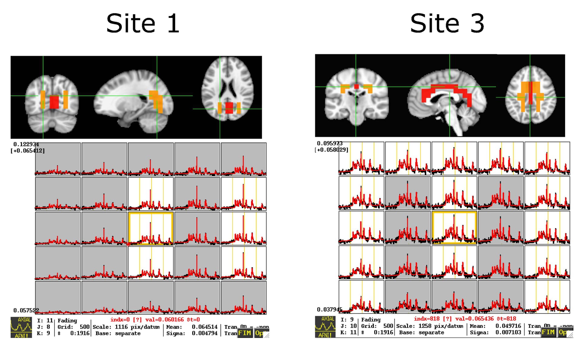
This multicenter study complied with the regulations of the local institutional Human Ethics Committees. 5 healthy volunteers were scanned at each site after giving written informed consent. Data were acquired using 3T whole-body magnets manufactured by Siemens (2 Prisma 3T systems and mMR (PET-MRI) system, Siemens Healthcare, Erlangen, Germany). The contributing institutes and corresponding scanner configurations were:
Purdue University, USA, Prisma Scanner, Dual Tuned Surface Coil, RAPID Biomedical
University of Bergen, Norway: mMR scanner, Dual Tuned Quadrature Head Coil, RAPID Biomedical
Goethe-University, Frankfurt, Germany: Prisma scanner, Dual Tuned Quadrature Head Coil, RAPID Biomedical.
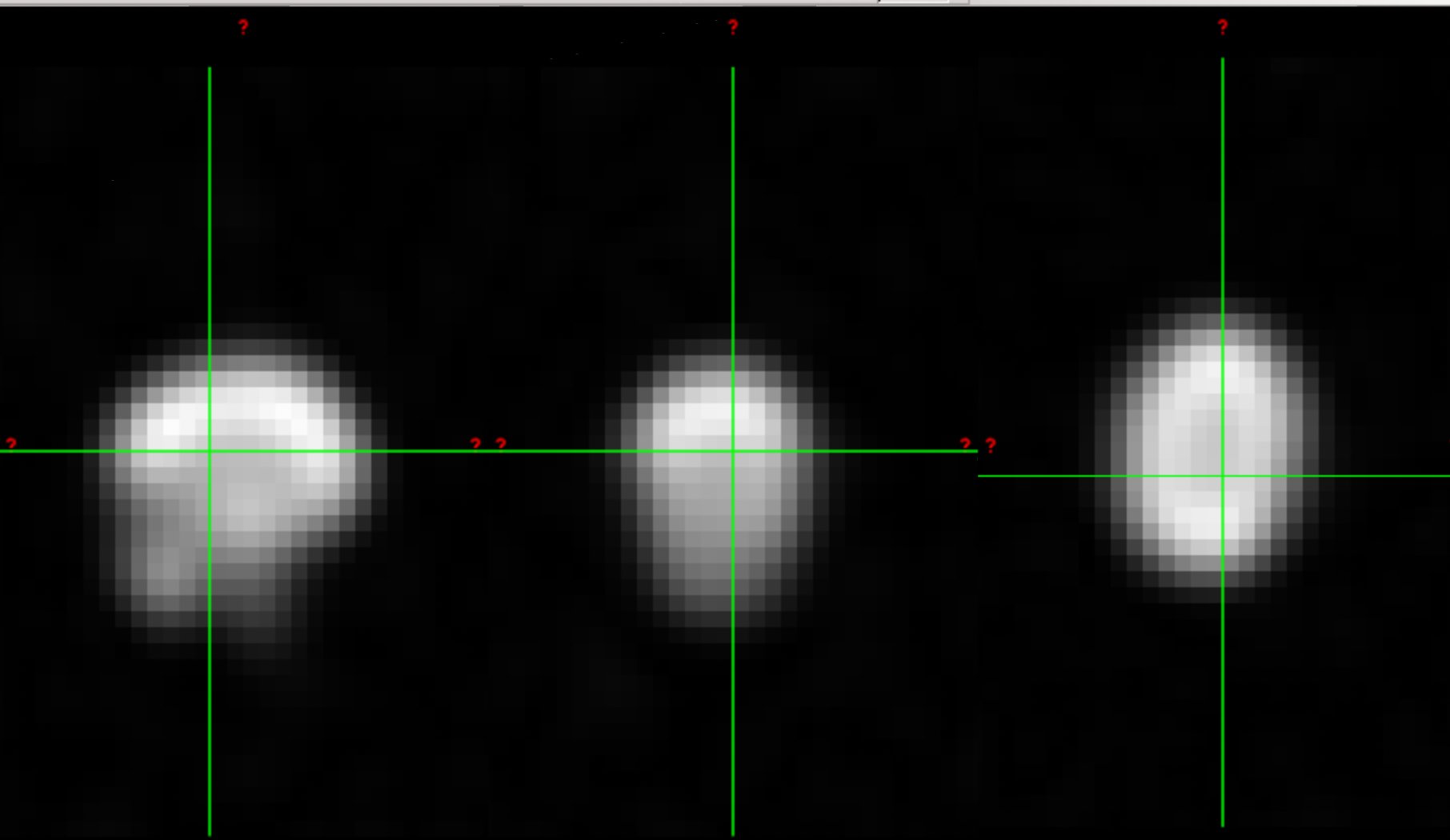
White and gray matter ROIs in occipital and motor cortices are defined due to different coil setups. The Purdue site used a surface coil with a brain coverage limited to the occipital cortex . Confidence intervals were calculated for each site and ROIs for the metabolite ratio of ATP/PCr. Confidence intervals for Z (0.95)-score in the occipital cortex for Purdue University are 9.281 ± 0.7871 and 9.029 ± 1.311 for gray and white matter ROIs, respectively. For the Goethe-University, Frankfurt, the values are 11.41 ± 1.183 and 10.21 ± 1.164, and for the University of Bergen, they are 10.83 ± 1.358 and 12 ± 3.559 for gray and white matter ROIs, respectively. Confidence intervals for Z (0.95)-score in the motor cortex for the Goethe-University, Frankfurt are 11.97 ± 1.089 and 11.04 ± 1.139 for the gray and white matter ROIs. For the University of Bergen, the values are 10.04 ± 1.388 and 9.555 ± 1.123, respectively.
uzay emir Purdue University
Ulrich Pilatus Goethe University Frankfurt
Seyma Alcicek Goethe University Frankfurt
Alexander Richard Craig-Craven UNIVERSITY OF BERGEN
uzay emir Purdue University
Ulrich Pilatus Goethe University Frankfurt
Seyma Alcicek Goethe University Frankfurt
Alexander Richard Craig-Craven UNIVERSITY OF BERGEN
“after 40 years, modern technology means clinical 31P MRs is finally feasible”
The ISMRM Challenge Committee Feedback
The judges would like to commend both sub-teams for taking on this Challenge! We’d like to provide you with some suggestions for future reproducibility efforts. To establish objective criteria for success in the preregistration (e.g. mean difference below a certain threshold) would improve the final evaluation of the effort. To eliminate any variability in a subsequent reproduction, we’d suggest compiling and releasing a processing pipeline and releasing the data acquired during reproduction. On the overall, this was a good reproduction effort!
Congratulations to the team,31P MRS Reproducibility got 2nd place. We will release the data and findings once we submit the full report for publication.
if you are interested in 23Na-Sodium MRI MSK and/or brain challenge, please drop me a message.
Multi-site feasibility and reproducibility study on UTE 3D phosphorous MRSI using novel rosette trajectory (PETALUTE)
we show that UTE 31P 3D rosette MRSI acquisition, combined with compressed sensing and LCModel analysis, allows fast, operator-independent, high-resolution 31P MRSI data to be acquired at 3T. Given the availability of 3T MRI scanners, the UTE 31P 3D rosette MRSI approach may have wide application in both clinical and research settings.