Single Shot Excellency is Key for fMRS

Hi All,

Last week a group of people interested in fMRS met via Zoom, with many thanks to @AnoukSchrantee and @admin for organizing!

During this meeting, @uzayemir made the comment that for fMRS “single shot excellency is key”. To me, this idea sparked many questions about how to optimize acquisition methods for fMRS applications. So I’m curious to hear whether the following ideas have been tested for fMRS? If not tested, do people feel that these would not greatly impact the ability to achieve single shot excellency?

  1. Cardiac synchronization (the brain does pulsate with the wave of arterial pressure)
  2. Respiratory synchronization (at 7T it has been shown to change the B0 shim in the brain)
  3. No phase cycling, instead relying more heavily on outer volume saturation
  4. Testing different chemical shift directions for each gradient direction to reduce outer volume signal
  5. Additional B0 shim coils or dynamic shim updates
  6. Placing dielectric pads outside the head to improve B1 homogeneity

… More?

Thank you all for your thoughts!
-Erin

2 Likes

Hi @erin.macmillan,

Thanks for opening this thread. Some really interesting suggestions here that matter for pretty much any MRS acquisition.

  • Any form of physiological synchronization (cardiac or respitatory) will add variability to shot-to-shot-TR, introducing T1 weighting that is practically impossible to disentangle from any functional change in metabolite levels. I guess respiratory sync would be less problematic, assuming 10 breaths per minute (every 6 seconds), but if you’re hunting for 1-2% functional changes, even that may be too much? Someone should make a study about that. :wink:
  • Another item for your list is optimized crusher configurations and phase cycling.
  • Reliable high-quality OVS seems really critical to single-shot excellency, but I guess it’s also really hard to get it to be reliable and high-quality (@richardedden always says that every pulse you add to a pulse sequence increases the chances of things going wrong). I like to believe that careful optimization of chemical shift directions is the prerequisite, a robust OVS and crusher setup should get you reasonably close, and dynamic updates to f0, shim, localization, OVS etc. should help the entire thing across the finishing line in terms of stability. There are a bunch of sequences out there now that are inching closer toward those goals (@VincentBoer for example).

TLDR: I think it’s super hard to get to single-shot excellency, particularly with stock sequences, but I like to believe that it can be achieved with great attention to detail.

Cheers,
Georg

1 Like

Hi Erin,
We have tried a couple of options that you suggest. As I remember you’re a Philips Clinical Scientist, so I’m curious to hear what your experience is.
When setting up fMRS at our Philips 7T, we used the dynamic loop to be able to synchronize the MRS sequence to the task, and at that time is was not possible to combine the dynamic loop with phase cycling - this really didn’t result in very nice spectra. In the new software release this was solved, and we’ve been using phase cycling ever since - which is very effective in getting rid of some artefacts, but perhaps not ideal for fMRS acquisitions.
With respect to using chemical shift directions, we have tried that as well and that really makes a difference what you choose, particularly for voxels near the skull. We then also apply OVS, but the efficacy of those can be variable, in my experience. So I’m interested in suggestions on how to improve this on Philips 7T.
I agree with Georg that the optimized crusher schemes (that still allow short TEs) will be very important - and an optimal combination with the OVS would be great.

Best,
Anouk

1 Like

Hello All,

I like the title :slight_smile:.
In general, we are trying to achieve artifact-free MRS data. For me, the inspiring/legendary study is Ivan Tkac’s 2001 work at 7T. Later on, those techniques were transferred to the clinical scanner (Siemens) by me and Edward J. Auerbach. This was followed by implementation on Philips (@VincentBoer, multi-site 7T work), and then @noeskera and I worked together to translate them to the GE when I was in Oxford.

Ivan also provided a recipe for this which I think it is the green book of MRS

In that paper, there are fantastic figures. The first one is illustrating the importance of single-shot data.

The second one is what it should like if you reach excellence.

This is what we desire. however, as you all highlighted it is not always achievable since each ROI has a different condition due to anatomical location and its interactions with MR. These imperfections can be tolerable for clinical studies.

However for fMRS and I think it will be wise to put the bar higher for a successful fMRS study from an MRI physicist’s point of view(I do not mean it will support the hypothesis of the study).

I absolutely agree with all the aforementioned suggestions. In addition to those here are my tricks (Ivan’s tricks).

Before starting any neuroscience-driven study (any physiological intervention MRS study is an fMRS), what I do is to test the sequence with subjects for specific ROIs and try to see possible problems in the spectra. I also record the shim values of problem spectra. For shimming my Suggestion will be not use FASTMAP for higher-order shims instead i have been using GRESHIM + FASTMAP (only for first-order shims). See below for the unsuppressed water due to the higher-order shims. I used this sequence during all my testings before starting any study at 7T

Then, I try to generate the problem spectra on the phantom using those shim values. Then I played with either the orientation of the Voxel (this is the easiest way but you need to make sure you are using the same orientation for all subjects, see @admin response) or crusher gradients. You can see above, you can send the unsuppressed water signal away from your actual FID using the crusher gradient amplitude in this case.

Then you will have an artifact-free/minimized study (See publications from Charlotte Stagg, Holly Bridge, and Zoe Kortzi’s groups in which I used these steps). Again, I am highlighting no-phase cycling reliance. Of course, it will minimize the problems and good to have but for fMRS studies, we need to be careful about its benefits.

I always try to get the best possible data since it will ease my life during post-processing and quantification (i really do not like to scratch my head about what went wrong during the acquisition.)

3 Likes

I love this post, @uzayemir. Are you taking any specific precautions with respect to the crusher amplitudes? I’m looking to implement DOTCOPS in our Philips systems soon, but I wonder about the benefit vs. a proper OVS.

The other thing I’ve been meaning to ask you about: do you ever see phase-cycle-dependent modulations of the lipid/MM area? I’ve seen extreme cases where the spectrum dips down into the negative between 0.9 and 2 ppm. I guess this can be modeled by flipping the MM/lipid basis functions on a case-by-case basis, but as you say, I’d rather not have to worry about it at all.

Ok, The reason i highlighted Ivan’s work: I only tailored them to work with B1 shimming and/or Nova coil. Once you setup these things, it works in almost every brain region without having any problem. So i need to highlight this one more time, the recipe is on the following paper.

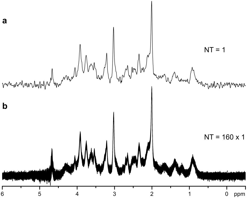
“Good localization performance and excellent water suppression resulted in highly reproducible single-scan spectra, without any significant contamination by unwanted coherences. This makes the sequence much more robust in comparison to sequences that require phase cycling to subtract unwanted coherences. When depending on phase cycling for the elimination of such signals, instabilities during an experiment, e.g., physiological motion of a subject, can result in subtraction errors and insufficient suppression of unwanted signals.To achieve the highest possible spectral resolution, elimination of all effects that unnecessarily increased the line width was desirable. Physiological motion, such as breathing, can cause periodic frequency variations up to ±2 Hz at 7 T. In addition, small random motions of the head during the study can induce phase fluctuations. Both effects were easily eliminated when single-scan data averaging was used (Fig. 2).”

There are two problems with OVS. One is it increases the SAR. But if you are using VAPOR then you do not need to worry about TR so it is not a problem anymore. The other one is the distance between OVS slabs and VOI. Ivan set this as 7mm and I tested them anything less than 7mm will result in signal suppression. I did have the illustration regarding this but I could not find it. Now if your lipid signal falls in between this region, then your VOI profile will bring it back to your spectra. This is a problem when you are working on occipital cortex ROI for fMRS. DOTCOPS or the following paper might solve the problem. But to be honest there is not any problem once you do shim and place the voxel very well.

https://onlinelibrary.wiley.com/doi/full/10.1002/mrm.26213

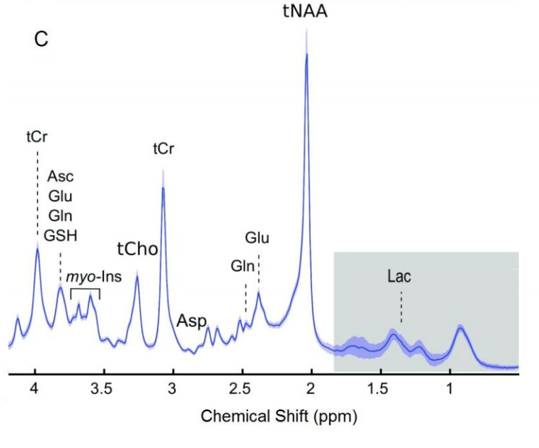
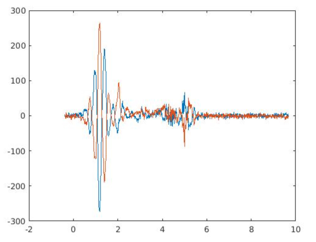
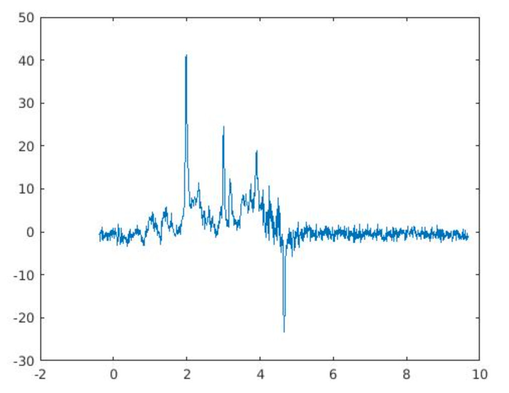
In my first OxfordWIN/fMRIB fMRS paper, we did have the problem of negative/positive peaks between 0.9 and 2 ppm, which was not terrible. Since I did not have the luxury of using CMRR hardware options, I was limited to standard hardware. Thus in the following paper, we used a limited ppm range.

https://www.cell.com/current-biology/pdf/S0960-9822(15)00434-0.pdf

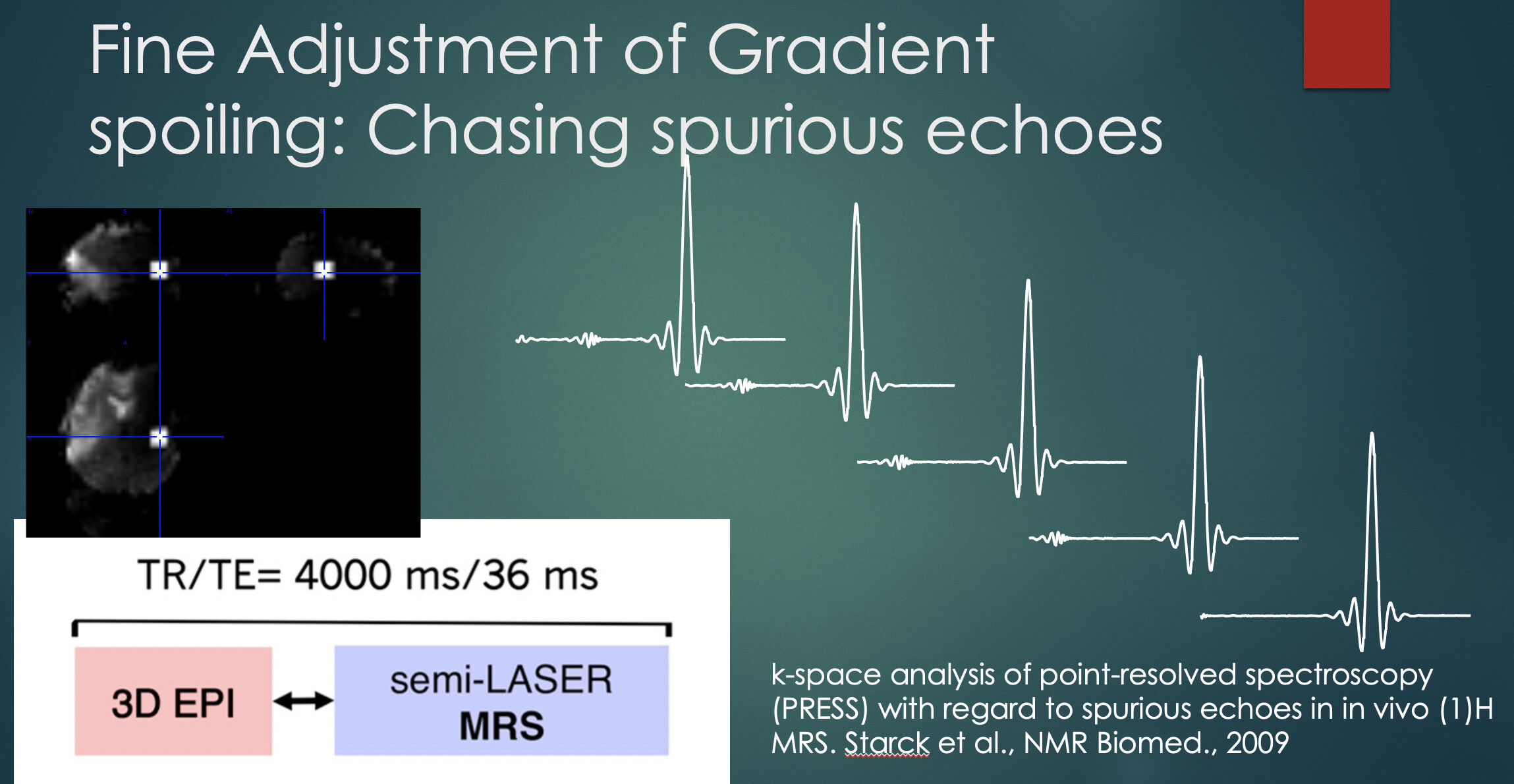
But I was not happy about this and I spent quite some time on RF pulse orders, gradients schemes since there was and is still a lot of demand at OxfordWIN/fMRIB for fMRS works. Then I reshuffled everything. CMRR spin-echo adiabatic (SEAD, , semi-LASER) sequence was using excitation in the X direction and their VAPOR-OVS partners (crusher gradients and pulses together). I realized with the NOVA coil this problem is originating from this order. then i change the orientation from Transverse to Coronal which resulted in a very reproducible spectral pattern in almost every brain region.

https://www.nature.com/articles/s41467-019-08313-y#MOESM1

Thus the problem you highlighted was not there anymore maybe a little bit in the ACC voxel.
I have done these in 2013/4 and I have not run any fMRS study and analyze them by myself. Thus Another trick to having a successful fMRS study is having excellent collaborators(Betina, Hellen, Zoe, Charlie, Holly… ) who understand/value MRS very well. Also, radiographers should also be on this team.
For instance, I have not acquired, analyzed, and reported any of these fMRS studies, I only looked at the results at the final stage. This way I minimized the possible bias that might originate from me. So most of them are blind design studies for me and we identify the analysis parameters in advance. Also, I have not acquired those data, this also minimizes any biases. For this, I have to thank OxfordWIN/FMRIB radiographers since they trusted me and followed the steps I suggested.

Overall, I think special thanks go to Ivan’s works.

3 Likes

Thanks for putting this online, really useful discussion, and thank you @uzayemir for the important technical points and links!

Martin

1 Like

Hi @erin.macmillan having the ability to get a TTL pulse out for each shot is something I think that should be added to your list. This allows time locking of the spectra to the stimulus presentation. Matthew Clemence once wrote a patch for us to do this, and so he may be able to help.

Hi @PGMM and @erin.macmillan,
At the 7T I think it’s now incorporated in the latest software version that you can use the TTL pulse to trigger each NSA and/or dynamic. You have to take into account when the trigger occurs though if you want to time-lock to your stimulus. If I remember correctly it’s at the start of the WS module (if you want to know for sure I can look it up), so then you have to time your stimulus such that you would give the first 90 degree pulse of your sequence at the moment you expect the Glu/GABA to be at a supposed peak. I have some e-prime examples of such a task. I would be interested for the patch to do this on 3T as well:).

1 Like

Very nice explanation, thanks @uzayemir! If you happen to find that figure on the OVS signal suppression I would be very interested - we’re using OVS for a lateral occipital cortex voxel, because indeed, we have quite disturbing lipid contamination. Even then, we use phase cycling, but indeed, I’d rather not have to deal with it.

For those wondering how that would look without phase cycling in an extreme case;
These are two subsequent averages (apologies for the reversed ppm axis) acquired with phase cycling:

Yet, if you average these you get this spectrum, so in this case the phase cycling is certainly necessary. However, this can give issues in the spectral registration if you don’t look at the individual averages and identify these artefacts.

Hello @AnoukSchrantee ,

i am digging into my hard drives, it was a lab report when I was at CMRR. Once I found it, I will share. Nevertheless, if your design is relying on timings like even related fMRS, I would emphasize again

“When depending on phase cycling for the elimination of such signals, instabilities during an experiment, e.g., the physiological motion of a subject, can result in subtraction errors and insufficient suppression of unwanted signals.”

We did an experiment with Helen Barron similar to your aim,

image

This is an event-related simultaneous fMRS-fMRI study, we tried to minimize all the artifacts. This allowed us to use all LCModel metrics during our analysis. For instance, any concentration changes can also be monitored by cLRBs.

image

so your extreme example has two problems, water signal coming from outside of your voxel due to higher-order shims and bad localization. The final spectrum is looking clean but the MM pattern has detoriated. The problem with this is how this deterioration is reproducible (I would be happy as long as it is reproducible). However, It will change with the number of averages and/or different subjects/ROIs. So your estimation of metabolites/CRLBs will not solely reflect task orignated metabolite changes instead it will be a mixture of phase-cycling and shifted water resonances.

Uzay

2 Likes

Dear @uzayemir,
Thanks very much! In this case we got rid of this major artefact by choosing a different orientation for the 90 degree pulse. Actually, for these experiments we use longer blocks, so the stimulus is not time-locked to the onset of the pulse, and so maybe in this case it’s a bit less disastrous. But it’s certainly something to look into for future experiments!
Best,
Anouk

2 Likes

Wise decision.

Just make sure you used the same orientation for all subjects, otherwise, you will have different chemical shift displacements.

Uzay

2 Likes

Wonderful thread… lots of interesting stuff here. @uzayemir : I am intrigued by your comments about gradients and the k-space reference (which I am yet to grasp)… are the out-of-voxel problem echoes gradient echoes (and not spin echoes as I had always assumed)?