Time Constrained PRESS & MEGA PRESS acquisition from same region

Hello everyone! I’m immensely grateful to this community for the generously shared knowledge! I apologize in advance if the following questions are basic!

I am new to the acquisition of PRESS (Glx) & MEGA-PRESS (GABA+). I plan to use Osprey to analyse and report PRESS and MEGA-PRESS data acquired from the same anatomical areas based on the recommendation given here.

I am acquiring spectroscopy data from cooperative clinical patients with severe symptoms in a hospital setting. We are allowed limited time in the scanner (total scan duration not to exceed 55 minutes), including several compulsory protocols (21 minutes, not to be modified).

Within the limited time left (34 minutes approx.), based on our clinical intervention, I wish to acquire single-voxel PRESS and MEGA-PRESS data from two anatomical sites: left dorsolateral prefrontal cortex (dlPFC) and left temporoparietal junction (TPJ). We are using Philips 3T Ingenia CX system v5.8 with a 32-ch head coil (I didn’t find an option to export protocol as a pdf!).

Based on the extensive feedback in this thread and this reference, we have come up with the following sequences. Given the time crunch, I need advice on optimizing acquisition parameters.

  • PRESS SEQUENCE FOR

    • Left dlPFC

      • voxel size: 20x15x20 mm=>6ml

      • Total Averages=64 [16 (No. of Signal Averages) x 4 (Dynamics)]

      • TR=3000 ms / TE=68 ms

    • Left TPJ

      • voxel size: 20x20x20 mm=>8ml,

      • Total Averages=64 [16 (No. of Signal Averages) x 4 (Dynamics)]

      • TR=3000 ms / TE=68 ms

  • MEGA-PRESS SEQUENCE

    • Left dlPFC :

      • voxel size: 32x27x32 mm=>27.64ml

      • Total Averages=144 [16 (No. of Signal Averages) x 9 (Dynamics)]

      • TR=2000 ms / TE=68 ms

    • Left TPJ : voxel size:

      • 30x30x30 mm=>27ml

      • Total Averages=144 [16 (No. of Signal Averages) x 9 (Dynamics)]

      • TR=2000 ms, TE=68 ms

  • Short TE scan for

    • Left dlPFC

      • Voxel size: 32x27x32 mm=>27.64ml

      • Total Averages=32

      • TR=2000 ms / TE=30 ms

    • Left TPJ

      • Voxel size: 30x30x30 mm=>27ml

      • Total Averages=32

      • TR=2000 ms / TE=30 ms

  • Water Un-Suppressed scan for

    • Left dlPFC

      • voxel size: 32x27x32 mm=>27.64ml

      • Total Averages=32

      • TR=2000 ms / TE=68 ms

    • Left TPJ

      • voxel size: 30x30x30 mm=>27ml

      • Total Averages=32

      • TR=2000 ms / TE=68 ms

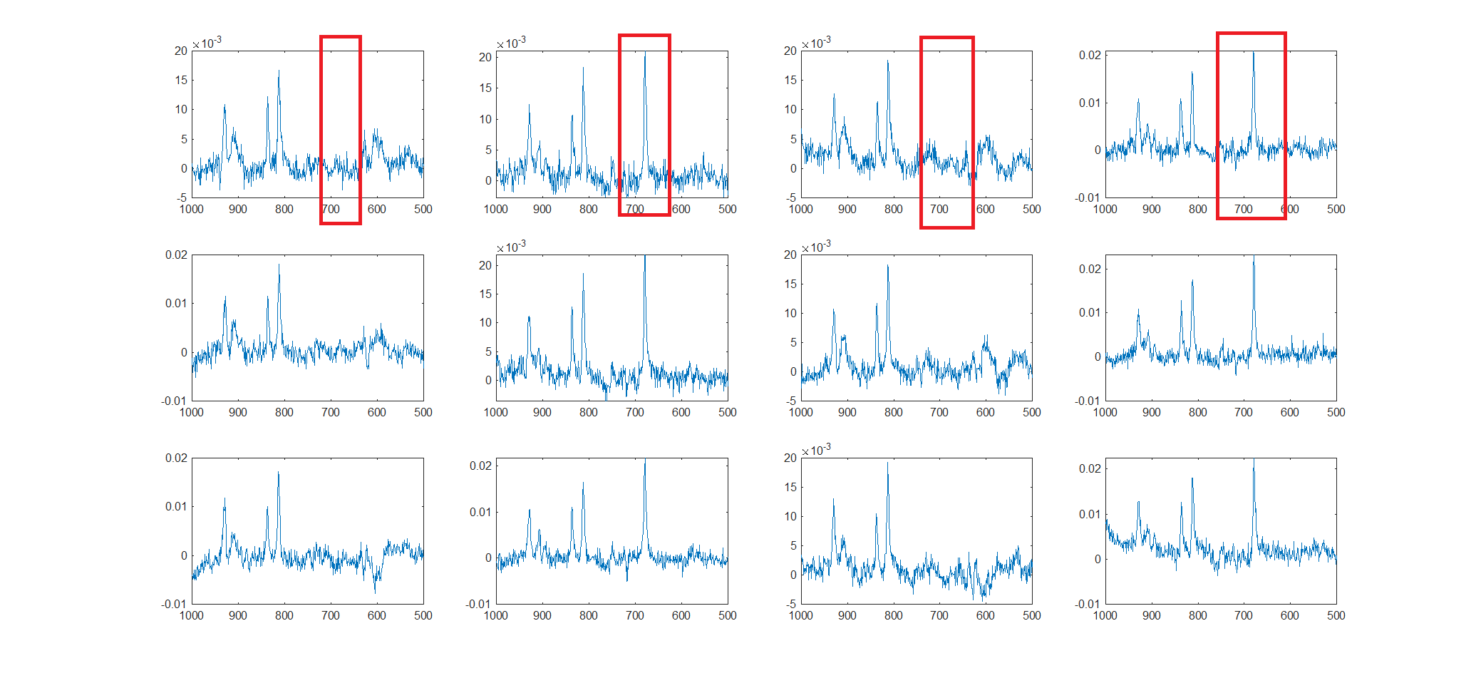
I have attached the concatenated exam cards for all these scan sequences as a single document. The compulsory protocol includes T1 weighted imaging (TR=6.5ms / TE=2.9 ms; exam card included as attachment). I will be using this for in-voxel tissue segmentation.

Anushree_Bose_EXAM_CARDs.txt (27.9 KB)

We prefer to acquire PRESS over MEGA-PRESS.

As I have limited time to dedicate to spectroscopy (34 minutes) in a 55-minute scan, any advice on what to keep and chop is truly appreciated! A few questions:

  1. What matters more for MEGA PRESS: acquiring more signal averages or accommodating a water un-suppressed scan and short TE scan? Is there a trade-off? Recommended number of averages is 300+, but we are acquiring 144! due to time constraints as we are accommodating Shoet TE scan and water unsuppressed scan.

  2. Should TR be kept same across PRESS, MEGA PRESS and Short TE? Is it okay to use the same Short-TE scan (TR=2000ms/TE=30ms) for both PRESS (TR=3000 ms/TE=68 ms) and MEGA PRESS (TR=2000 ms /TE=68 ms) if targeting the same anatomical areas?

  3. Is it correct or incorrect to vary the voxel sizes between PRESS, MEGA PRESS and Short-TE if targeting the same anatomical region? At present, I have kept the voxel sizes as same for Short-TE and MEGA PRESS but smaller for the PRESS sequence.

  4. Are start-up scans (instances where complete pulse sequences is run without acquiring data in order to establish steady state) or dummy scans mandatory? If yes, are 4 ‘start-up scans’ enough?

If the time limit is too constraining to acquire sufficient averages along with Short-TE / Water Unsuppressed scans, I am open to dropping either left dlPFC or left TPJ. However, given the clinical context and relevance of the study, retaining both sites is desirable.

Looking forward to improving our efforts with your input!

Thanks and regards,

Anushree

Hi Anushree,

Some thoughts (similar to the ones I’ve shared in this thread yesterday) below.

  • Most importantly: You don’t need a separate TE = 68 ms PRESS scan if you’re already running a TE = 68 MEGA-PRESS scan. The MEGA-PRESS edit-OFF will be identical to the TE = 68 ms PRESS scan (assuming that you put the edit-OFF editing pulse far away from anything, like at 8 or 9 ppm). Getting rid of this separate scan will free up transients that you can spend on the MEGA-PRESS. I don’t understand why this particular scan is the only one that is targeting a (massively smaller) volume. This would quite likely not get enough SNR to be viable (SNR drops linearly with measurement volume).
  • Speaking of MEGA-PRESS: Get as many MEGA-PRESS transients and as large a voxel as you can. 144 transients is awfully short.
  • I would probably try and keep TR consistent across all scans.
  • I would also keep the voxel sizes consistent across all scans.
  • The reason is that you can use the same short-TE water reference scan for quantification for both the short-TE PRESS and the medium-TE MEGA-PRESS acquisitions.
  • You can probably reduce dummy/start-up scans to 2 instead of 4. Saves a few seconds.
  • You don’t have to set up separate water reference scans. Philips can acquire these for you automatically. Just set PostprocSpectral Correction to yes for all of your scans (MEGA-PRESS and short-TE PRESS) and specify the number of transients in the next line. These will be exported as _ref.sdat/spar.
  • You definitely don’t need 32 transients for your water reference data. 1-4 will absolutely suffice.
  • If you have VAPOR as a water suppression option, go for that one. If not, go for any other option (e.g., MOIST) than excitation.

Here’s what I’d set up for each region (left DLPFC 32 x 27 x 32 mm3; left TPJ 30 x 30 x 30 mm3) if I had 34 minutes to burn:

  • Short-TE PRESS (TR/TE = 2000/30 ms)
    • 64 transients
    • 1 water reference transient
    • total duration: 2 sec * (64 + 1 + 2) = 134 sec (2:14 mins)
  • MEGA-PRESS (TR/TE = 2000/68 ms)
    • 320 transients
    • 1 water reference transient
      • ADDENDUM: I can’t quite recall how the product MEGA sequence does it, but I believe it can do an interleaved water frequency update similar to what we built a few years ago. I can probably find that out (tagging @sganji in the hope he might see it)
    • total duration: 2 sec * (320 + 1 + 2) = 646 sec (10:46 mins)
      • slightly longer if you increase the number of water reference transients

So per region you’ll be clocking in at around 13 mins pure measurement time. You’ll want to allow for proper preparation in each region (calibrations, shimming, water suppression optimization etc. take about a minute to a minute and a half). And you always, always, want an extra few minutes in your protocol if things go wrong and you need to restart a scan (say, you get a bunch of lipids in your signal and you need to reposition your voxel. Absolutely common in dlPFC).

This protocol would leave you with very good quality data and give you everything you need and, most importantly, it would not put you under the intense pressure of not being allowed the tiniest failure. (This can make all the difference between OK-ish data and really excellent data).

Cheers,
Georg

1 Like

Some bonus thoughts after checking your protocol txt file:

  • I’d set the number of points to 2048 (keep 2 kHz spectral width). Gives you a few extra data points per ppm. Keep consistent for all MRS scans.
  • Acquire all scans for each region consecutively, then turn over to the other region. Don’t alternate between regions (it will trigger re-shim and re-calibration). If you give the two scans of the same region the same geometry name, it should lock their localization together (but always double-check!!).
  • For the first MRS scan in each region, set Preparation phases to full; the second one can just be set to auto.
  • For MEGA-PRESS in particular, you will want to optimize the slice-selective gradient orientations to minimize out-of-voxel echoes (see thread here).
    • There is a caveat to this: since you’ll be operating in cortical regions very close to the skull, you’ll want to make sure you’re not getting lipid contamination. This can be achieved by moving the voxel a few mm away from the skull or by making sure that the chemical shift displacement (denoted by the second box; set PostprocShifted metabolite displayed to Lip/Lac and determined by the slice-selection gradient polarity settings Chem Shift Dir AP/LR/FH) puts the area where the lipids towards the inside of the brain. Again, make sure these settings are completely consistent for all of your scans.
  • Make sure that all of your more generic parameters are identical between MRS scans of the same region (e.g. VOI orientation once you have determined the best one).
  • Very specifically, make sure that the GABA-editing settings are exactly identical between regions. The bandwidth is set to 80 Hz for one and to 100 scan for another. Choose the lowest possible value here (it’ll be around 80 Hz for TE = 68 ms).
  • You can also reduce the shim size setting - I think it’s usually sufficient to add 5 mm to the voxel volume in all directions.
  • I’d set PNS mode to the highest possible value.
1 Like

Thank you so much, Dr. Oeltzschner!

I am immensely grateful for this detailed, point-by-point response. I will revise the protocol with the help of our MR technicians to incorporate the changes you advised.

I am just starting with spectroscopy, and your responses are beyond helpful!

1 Like

Let me shed some light on the Philips product MEGA-PRESS implementation.

Default protocol named “SV_MEGA_GABA” has the following parameters"
(located in the examcards tree at Philips > Head > Spectroscopy_1H > Brain)
TE = 68ms; TR = 2000 ms; SW = 2000; Number of points = 1024 Basing/MEGA pulse = MEGA; Window = 150 Hz; MEGA ON offset ="GABA(1.89 ppm)"; MEGA OFF offset = "default" (7.46ppm); shim = PB-Auto; PB-auto order = first; Water suppression = "excitation"; window (Hz) = 140; second pulse angle = 300; window offset (PPM) = 0; NSA = 16; Phase cycles = 16; Startup acquisitions = 0; freqency stabilization = voi (dynamic); Dynamics = 9 (individual); Plan scan metabolite = Creatine (3.02 ppm); Shift metabolite displacement = NAA (2.01 ppm)

Please note that PB-auto order should be set to SECOND at 3T

Now, MGEA sequence looping proceeds as follows:

for d = 1 : numberOfDynamics
	Performs one unsuppressed Water acquisition
	--Updates the F0 (frequency for next set of NSA)
	--Used for eddy current correction (zero/first order correction)

	for knsa = 1 : numberOfAverages (NSA)
		EDIT ON  with PhaseCycle(knsa)
		EDIT OFF with PhaseCycle(knsa)
	end
end

If one uses the above protocol, they will end up with 297 FIDS [ 16 (NSA) * 2 (On/OFF) * 9 (Dynamics) + 9 (water ref scans) ]. If exported it as SPAR/SDAT data, one will have 288 FIDS in *raw_act.SDAT and 9 in *raw_ref.SDAT file. If exported as Classic DICOM then 297 spectro DICOM files will be created.

Best,
-Sandeep

2 Likes

Thanks @sganji, this is great, so it’s indeed an interleaved water frequency update, cool.

Compared to the default Philips MEGA protocol, the editing pulse bandwidth (default 150 Hz) needs to be lowered as much as possible to maintain the best possible frequency selectivity (as I described above, it should be about 80 Hz)

1 Like

Yes, I spoke with @richardedden regarding this. I’m currently in the process of updating the protocol within the Philips default database. Please be aware that these updates require time to propagate to end users.

Until then, I recommend users to use the below protocol on Philips 3T scanners (note the default protocol is already mentioned in the above post).

Modified protocol for MEGA-GABA on Philips 3T
TE = 68ms; TR = 2000 ms; SW = 2000; Number of points = 2048
Basing/MEGA pulse = MEGA; Window = 88 Hz; MEGA ON offset =“GABA(1.89 ppm)”; MEGA OFF offset = “default” (7.46ppm);
shim = PB-Auto; PB-auto order = second
Water suppression = “MOIST”; window (Hz) = 140; second pulse angle = 300; window offset (PPM) = 0;
NSA = 16; Phase cycles = 16; Startup acquisitions = 0
freqency stabilization = voi (dynamic)
Dynamics = 10 (individual)
Plan scan metabolite = Creatine (3.02 ppm)
Shift metabolite displacement = Water (4.68 ppm)

I highlight the ones that needs to be particularly changed/checked when making the modified protocol on the scanner.

Note:
-Basing (editing) pulse bandwidth changed from 150 Hz to 88 Hz
-Users can choose to perform Water suppression = “VAPOR”, if they have the VAPOR software license
-Users can choose to perform shim = PB-Volume, if they prefer

1 Like

Dear Dr Oeltzschner @admin, your comments are immensely valuable!

Thank you for clarifying the Philips MEGA PRESS protocol, Dr Ganji @sganji!

We are still struggling to optimize the ‘right’ setting for our spectroscopy protocols in the Philips Inegia scanner with 32 channels.

I have a few follow-up questions! It would be great if anyone could give further clarification.

Question 1

Any suggestion on how to split the MEGA PRESS, TE=68ms SDAT/.SPAR files correctly to extract MEGA PRESS “edit-OFF” data to use as PRESS, TE=68ms data for Glx analysis? We could not find the scanner setting/any other way to do this.

Question 2
Please advise on how to save raw data for PRESS. I am not sure if our current settings are optimal.

For our pilot recording of Short-TE PRESS (TR/TE=3000ms/35ms) sequence and MEGA PRESS (TR/TE=2000ms/68ms), we set PostprocSave raw data to ‘yes’ with

Option 1: MotionNSA (set to 64): The Chemical shift drift graph is not generated after running the Process Data in Osprey for PRESS.

MotionNSA (set to 144): The chemical shift graph is generated with adequate data points for the MEGA PRESS sequence.

Option 2: MotionNSA (set to 16) + Dyn/Angdyn scans (set to 4).
The chemical shift graph for the PRESS sequence is generated with only 4 data points (for each pre and post-spectral alignment).

MotionNSA (set to 16) + Dyn/Angdyn scans (set to 9 or higher)
The chemical shift graph is generated with adequate data points for the MEGA PRESS sequence.

Do we need a minimum number for NSA or dyn scans for an ideal frequency drift graph for PRESS? Why is the frequency drift graph display affected in PRESS but not in MEGA PRESS?

I have attached the outputs from the Osprey Process data step for the above-mentioned options from both PRESS and MEGA PRESS.
Anushree_Bose_Osprey_Process_Outputs.pdf (691.7 KB)

For Short-TE PRESS, Specifying only NSA doesn’t generate a frequency drift graph. Additionally, the spectra look averaged in both the NSA only & NSA + dyn scans options for PRESS. How do I save unaveraged, raw data?

Specific to the Philips scanner, I see the word transients mentioned in papers. Is there a setting where this can be specified? Specifying NSA with a higher number of dyn scans greatly increases the scan time. For example, 16 NSA + 20 dyn scans clocks over 23 minutes.

Question 3
I understand that if the parameters (TR, TE, voxel size, etc.) are kept consistent, MEGA PRESS ‘edit off’ data can be used as PRESS data without another independent PRESS acquisition. However, what is preferred/recommended for a reliable, high-quality Glx & GABA+ quantification from a clinical population involving repeated spectroscopy experiments targetting difficult brain regions like the left dorsolateral prefrontal cortex and left temporoparietal junction?

  • Short TE-PRESS (TR/TE=3000ms/35ms) with a smaller voxel size for Glx and a separate intermediate-TE MEGA PRESS sequence (TR/TE=2000ms/68ms) with a much larger voxel size for GABA+, or
  • A single MEGA PRESS sequence with identical parameters for both Glx and GABA+

As a beginner, I am conflicted from the literature review. Experts in the field, please share what you would do for your study in a similar context.

Lastly, immense thanks to this forum, and shout out to Dr Oeltzschner @admin for spreading incredible knowledge!

Cheers,
Anushree Bose

Question 1:
The order is Edit ON and Edit OFF. One can also quickly confirm this by plotting the data after FFT, you can see the NAA signal is not present in EDIT ON spectrum, while its there in EDIT OFF spectrum.

Question 2:
Please note that rawdata can mean many things depending on the context. SPAR/SDAT data are final reconstructed spectra (same data as in DICOM files). DATA/LIST can give you individual transients (excitations) and all the channels of the coil used, but then you will have to reconstructed data offline.

Now, the final number of FIDS in the SPAR/SDAT depends on the parameters of the acquisition of PRESS/STEAM
Example:

NSA		Dyn		No. of Transients 	No. of spectra in SPAR/SDAT
64		1		16*1=64		1
16		4		16*4=64		4
16		9		16*9=144	9

Now, for MEGA-GABA Philips implemented special reconstruction such that one can get all the transients.
Example:

NSA		Dyn		No of spectra in SPAR/SDAT for MEGA-GABA
16		9		16*9*2=288 (2 is for EDIT ON and EDIT OFF)
16		10		16*10*2=320 (2 is for EDIT ON and EDIT OFF)
8		10		8*10*2=160 (2 is for EDIT ON and EDIT OFF)

One transient means one TR (excitation + acquisition more specifically)

Question 3:
I will let Georg recommend, but if you can do the

Hope that helps.

1 Like

Thanks Sandeep, super great. To add:

Question 1:

You do not need to split anything if you analyze data with Osprey. It will automatically fit the edit-OFF spectrum (in the GUI, you can cycle through the different fits and quantitative results by clicking the ‘Spec’ buttons in the top left corner).

Question 3:

If you want to compare your results across acquisitions, you want the signal to come from the same location. I don’t think it’s good practice to change the MRS voxel size but stay in the same location.

In general:
For GABA at 3T, consensus is fairly broad that you will definitely need spectral editing.

For Glx/Glu/Gln, the picture is less clear. What we know from literature is that Glx/Glu estimates derived from the different techniques (PRESS vs. edit-OFF vs. edit-diff) do not agree very well. The most likely explanation is that the signals have different degrees of overlap with each other depending on the echo time and whether it’s a difference spectrum. The underlying macromolecular signal profiles will also be different between these three experiments, and it has always been a big challenge for modeling algorithms to properly separate the metabolite from the MM signals.

If you ask 20 spectroscopists, you’ll get 20 different answers for almost any question, and the question “what should I use to estimate Glx best?” is another one. I won’t give you an answer that I claim to be the definitive one. You’ll see a bunch of papers out there, but IMO none of them have provided convincing evidence in favor of one answer or the other.

1 Like

Hi @sganji
Thanks for adding this, it’s useful for others who may want to set up the Phillips MEGA-PRESS implementation as well. One question though - how do you set the MEGA pulse bandwidth to 88 - and still keep the TE at 68? When I do this on my system (3T Elition) the shortest TE it will allow is 78 ms? I’ve gone with a TE of 80 ms instead, as this is one of those TE’s that is supposed to give a “better” Glutamate fit. As Georg says, most of these suggested optimised Glu methods are optimised for reliability. Accuracy is another issue - and likely relies on the fitting being used as much as the acquisition.
Note, on the issue of differences between short TE PRESS and edit OFF PRESS - again this may be more to do with the peculiarities of the fitting software being used, than the acquisition technique.
@BoseA your best bet for Glutamate is to get a good short echo PRESS/semi-laser.

1 Like

Thank you so much for explaining the basics, Dr Ganji @sganji !
This thread is an excellent resource for Philips scanner users! Regards.

Thank you so much, Dr Oeltzschner @admin!
I am just beginning to scratch the surface here, and your explanations and advice have been beyond helpful! As I am starting out with spectroscopy, I will try Short TE PRESS first. As you have advised, keep the voxel size the same across acquisitions.

Thanks for the advice, Prof. Mullins @PGMM! Much appreciated!

You will have the change the Gradient mode so that crushers can be played within the TE = 68 ms.
Have you tried changing the Gradient Mode (on Contrast Tab) to “maximum”?

-Sandeep

Hi Sandeep,
I managed to get the TE down to 68 ms in the end, thanks for the suggestions.

Good. Let me know if you other questions.