Dear development team,
I have discovered that the voxel mask created in the coregistration step seems to be flipped left-right, see the attached pdf file. This will probably affect the obtained tissue fractions in the segmentation step and also the quantification based on water and tissue corrections but this needs to be checked carefully to make sure that the correct voxel position is used in the calculations.
/Greger
That’s indeed concerning, thank you for the bug report. Can you please let me know whether you used DICOM or NIfTI data for the co-registration, and what GE software version this is? I’m quite sure we got it right for Siemens and Philips, but Helge and I personally haven’t done a lot of GE processing, and the feature to co-register straight to NIfTI instead of DICOM has only been added recently.
Thanks for helping us make Osprey better, and happy holidays,
Georg
Essentially the voxel placements were aligned to the posterior gray matter and parietal white matter as indicated in:
Lin, A., Tran, T., Bluml, S., Merugumala, S., Liao, H. J., & Ross, B. D. (2012, September). Guidelines for acquiring and reporting clinical neurospectroscopy. In Seminars in neurology (Vol. 32, No. 04, pp. 432-453). Thieme Medical Publishers. DOI: 10.1055/s-0032-1331814
Would I need to specify the folder containing all of the associated dicom files? I have tried that but keep getting the error:
Error using spm_vol>spm_vol_hdr (line 80)
File “H:\PhD\RA-MRS\Jobfiles\ErrorChecks\sub-43\ses-01\anat” does not exist.
thanks for the update. So just to clarify the current state. The PDF that I posted was still not right? I did test it with the nifit you send me and a nifti generate with MRIcroGL and the voxel position matched.
It is relatively hard for me to figure out how for off the positioning is, as I did not run the study or have a real reference. Do you have the voxel positioning images generated on the scanner ( It is something like save images and spectra in the spectroscopy tab) or can you go back to the scanner to generate them? It would be much easier if I could see them or a screenshot/photo of the on scanner positioning.
DCM input is currently only possible for GE data. You could look into the coreg_p function to see how this is set up, using SPM to generate the nifti.
Hi. Just in case anyone else stumbles across this thread wondering why their data are flipped L-R… Osprey appears to be using NEUROLOGIC convention (L side of brain on L side of image) and NOT radiologic (L on R) convention.
I processed multiple datasets from Philips and GE, using T1s that were converted from dcm2nii using all of the same tools above (i mean all). And achieved the same result. Then tested this with data that I have with clear demarcation of L-R (e.g., unilateral lesions) and am consistently seeing things flipped L-R, but in the appropriate location relative to the anatomy.
Helge, I’m curious, is there a flag I can use to flip things into radiological convention for those of us who prefer that (and already have enough R-L confusion ?
Sorry to resurrect an old thread - but I am having a similar problem. I have run some data through both Gannet and Osprey, and it appears that the voxel is being flipped in Osprey.
I’ve attached the coreg output from both Gannet and Osprey - the voxel should be left ventral visual, which it is in Gannet, but not Osprey. As @jwisnowski asks, is there a flag to easily flip things in Osprey?
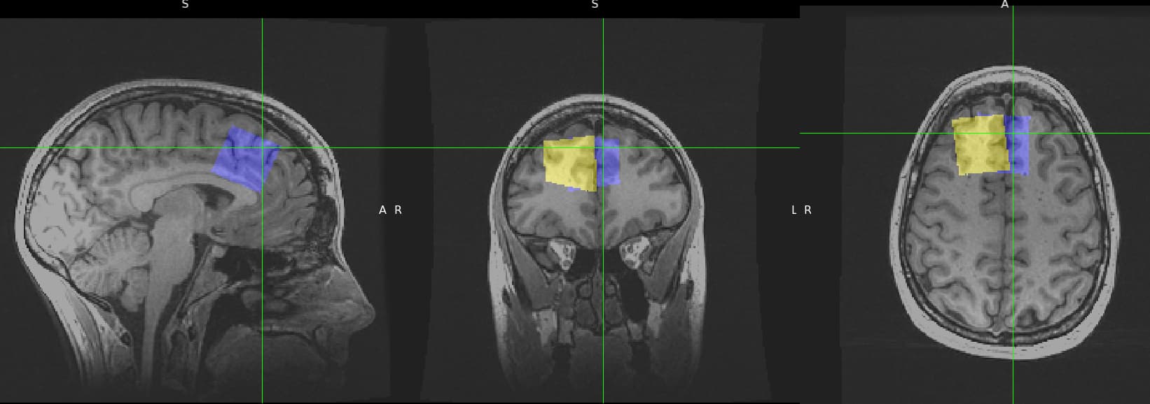
Adding to @mollysimmonite’s observation - I’m having weirdness with the CoReg function in Osprey on GE data, that has been converted to nifti (through Gannet’s routine for the most part I believe).
Pictured is the Gannet (blue) and Osprey (yellow) overlaid
I hope that this is really just about an LR flip. I’ll come up with an option to do this as this seems to be a common issue with GE MRS/imaging data depending on the NifTi conversion.
 ?
?